MICRON BIOIMAGING FACILITY
Access to advanced imaging technology
Including bespoke development systems
With expert guidance and support for your project

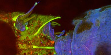
We help researchers apply cutting edge fluorescence microscopy technology to their research questions. See our range of microscopes here.
Find out how to access our cutting edge imaging technology including our super-resolution and development systems.

Wide ranging high profile publications from addressing key biological questions to development of new instruments and approaches.

Find out our latest events and news happening in the facility.
Access to all our fluorescence microscopy and image analysis educational tools.

The primary source of funding for our cutting-edge imaging research.
Principal Applicant: Ilan Davis
Co-applicants: Jordan Raff, Martin Booth, Yvonne Jones, Christian Eggeling, David Stuart, Kay Grunewald, Neil Brockdorff.

Funded by the Wellcome Institutional Strategic Support Fund and the John Fell Fund, these two innovative microscopy development projects, the 4Pi SMS & Microscopi, are aimed at very different areas of research.